Prof dr Marinko L Žuvela
Prof dr Žuvela operates on all types of hernias of the anterior and lateral abdominal wall through an open approach. It surgically resolves all preoperative complications of hernia (pinched – strangulated hernia) or postoperative complications of hernia surgery (recurrent hernia; mesh infection after hernia surgery; chronic pain after hernia surgery – inguinodynia).

Groin hernias (inguinal and femoral) are solved in different ways depending on the clinical findings. Small and medium inguinal and inguinoscrotal hernias are solved with the Lichtenstein technique, and femoral hernias with the plug + Lichtenstein technique under local anesthesia according to the principles of ambulatory surgery. The patient leaves the clinic 2 hours after the operation.
*Žuvela M. The modified Lichtenstein technique for complex inguinal hernia repair – How I do it. Acta Chir Iugosl 2011; 58(1):15-28.
*Žuvela M. Ambulatory surgery of abdominal wall hernias – Ten years of experience. 36th International Congress of the EHS, Edinburgh, Scotland 2014


He solves large inguinoscrotal hernias with his original method – “Rives technique performed through a direct inguinal approach” under local anesthesia, and depending on the local findings, the patient leaves the clinic 2 hours after the operation or spends 1 day in the clinic.
*Žuvela M, et al. Rivesova tehnika Direktan ingvinalni pristup u rešavanju velikih ingvinoskrotalnih i recidivantnih kila. Acta Chir Iugosl 2003;50(2):37-48.
*Žuvela M, et al. Modifikovana Rivesova tehnika u tretmanu recidivantnih ingvinalnih kila. Acta Chir Iugosl 2003;50(4):53-6.


Small and medium ventral hernias (umbilical, epigastric, Spigel’s, incisional) are solved with his original method – “open preperitoneal flat mesh technique” with the installation of a small mesh under local anesthesia according to the principles of ambulatory surgery. The patient leaves the clinic 2 hours after the operation.
*Žuvela M, et al. Spigelian hernia repair as a day-case procedure. Hernia 2013;17(4):483-6.
*Žuvela M, et al. Management of epigastric, umbilical, spigelian and small incisional hernia as a day case procedure: results of long‑term follow‑up after open preperitoneal flat mesh technique. Hernia 2021;25(4):1095-1101.
*Žuvela M, et al. Day case surgery of umbilical, epigastric and small incisional hernias – open preperitoneal flat mesh repair under local anesthesia. 29th International Congress of the EHS, Athens, Greece 2007
*Žuvela M, et al. Incisional hernia surgery as a day case procedure. 30th International Congress of the EHS, Sevilla, Spain 2008
*Žuvela M. Groin, umbilical, epigastric and spigelian hernias as a day case surgery. 30th International Congress of the EHS, Sevilla, Spain 2008
*Žuvela M, et al. Spigelian hernia repair as a day case procedure. 4th Joint Hernia Meeting of the AHS and EHS, Berlin 2009
*Žuvela M, et al. Ambulatory surgery of ventral and incisional hernias. 32nd International Congress of the EHS, Istanbul, Turkey 2010
*Žuvela M, et al. Lowcost technique for the management of all types of ventral hernias in ambulatory settings – safe and reliable procedure. 39th International Congress of the EHS, Wiena, Austria 2017
*Žuvela M, et al. Management of epigastric, umbilical, spigelian and small incisional hernia as a day case procedure: results of long term follow up after open preperitoneal flat mesh technique. 44th EHS Annual Meeting. Manchester, UK 2022


Large midline incisional hernias are solved with the Rives sublay technique with the installation of a large mesh under general anesthesia.
*Žuvela M, et al.. Rives technique (sublay) in the management of major incisional hernias. 2th International Congress of the EHS, Turin, Italy 2005
*Žuvela M,et al. Is the sublay repair a technique of choice for major incisional hernias on any location? 29th International Congress of the EHS, Athens, Greece 2007


Giant midline hernias – eventrations are solved by one of the techniques of component separation + mesh hernioplasty under general anesthesia.
*Žuvela M, et al. Possible solution for complex abdominal wall defects – various components separation techniques with or without nonresorbable mesh hernia repairs. 32nd International Congress of the EHS, Istanbul, Turkey 2010
*Žuvela M, et al. Eventration – complex abdominal wall defect. 35th International Congress of the EHS, Gdansk, Poland 2013.
*Žuvela M, et al. How to prevent intraabdominal hypertension and postoperative compartment syndrome following incisional or ventral repair and acute wound bursting – Various techniques of components separation with mesh augmentation. 36th International Congress of the EHS, Edinburgh, Scotland 2014
*Žuvela M, et al. Individual approach for patient with ventral eventration: various components separation technique with mesh augmentation. 38th International Congress of the EHS, Rotterdam, The Netherlands 2016
*Žuvela M, et al.. Individual approach for a patients with abdominal wall eventration – various componenets separation technique with mesh augmentation. 41st International Congress of the EHS, Hamburg Messe, Germany 2019
*Žuvela M, et al. Individual strategy for patients with abdominal wall eventration – different componenets separation technique with mesh augmentation. 42nd Annual Congress of the EHS, Barcelona 2020
*Žuvela M, et al. Individual strategy for patients with abdominal wall eventration -different components separation technique with mesh augmentation. 44th EHS Annual Meeting. Manchester, UK 2022


He solves large subcostal hernias – eventrations without signs of infection with his original method – “modified sublay technique for large subcostal hernias” under general anesthesia.
*Žuvela M, et al. The modified sublay technique for the management of major subcostal incisional hernia – long term follow-up results of 37 consecutive patients. Ann Plast Surg 2022; 1;88(2):212-218.
*Žuvela M, Galun D. Complex subcostal abdominal wall defect – Three variations of components separation technique combined in one procedure. 36th International Congress of the EHS, Edinburgh, Scotland 2014
*Žuvela M, Galun D. Combination of the three techniques in one procedure for management of complex subcostal abdominal wall defects. 1st World Conference on Abdominal Wall Hernia Surgery, Milan, Italy 2015
*Žuvela M, et al. The modified sublay technique for management major subcostal incisional hernia: Rives sublay technique with components separation technique. 39th International Congress of the EHS, Wiena, Austria 2017
*Žuvela M, et al.. The modified sublay technique for the management of major subcostal incisinal hernia. 41t International Congress of the EHS, Hamburg Messe, Germany 2019
*Žuvela M, et al The modified sublay technique for the management of major subcostal incision eventration. 42nd Annual Congress of the EHS, Barcelona, Spain 2020
*Žuvela M, et al. The modified sublay technique for the management of major subcostal incisional hernia: long-term follow-up results of 41 consecutive patients. 44th EHS Annual Meeting. Mancheste, UK 2022.


He solves large subcostal hernias – eventrations with signs of infection using his original method – “combination of three component separation techniques” under general anesthesia.
*Žuvela M, et l. The combination of the three modifications of the component’s separation technique in the management of complex subcostal abdominal defects. Hernia 2022. May 16. doi: 10.1007/s10029-022-02622
*Žuvela M, et al.. Complex subcostal abdominal wall defect – three models of components separation technique fussed in one procedure. 41thInternational Congress of the EHS, Hamburg Messe, Germany 2019
*Žuvela M, et al. Three models of components separation technique fussed in one procedure with mesh augmentation: management of complex subcostal hernia. 42nd Annual Congress of the EHS, Barcelona 2020
*Žuvela M, et al.. The combination of the three modifications of the component separation technique in the management of complex subcostal wall defect. 44th EHS Annual Meeting. Manchester, UK 2022


The simultaneous existence of 2 large ventral hernias (a midline hernia and a lateral hernia of any size) is solved by a combination of the sublay technique and the component separation technique + mesh hernioplasty with a large mesh under general anesthesia.
*Žuvela M, et al. The modified components separation technique – one stage solution for contaminated major midline and transrectal abdominal wall defects. 30th International Congress of the EHS, Sevilla, Spain 2008
*Žuvela M, et al. How to repair a lateral incisional abdominal wall hernia? 44th EHS Annual Meeting. Manchester, UK 2022


Parastomal hernias are solved with the technique of component separation + “kyhole” or Sugarbacker mesh hernioplasty with a large mesh under general anesthesia.
*Žuvela M, et al. Parastomal hernia repair: Modified Sugarbaker procedure with components separation technique vs modified Rives sublay with components separation technique. 38th International Congress of the EHS, Rotterdam, The Netherland, 2016
*Žuvela M, et al. Open parastomal hernia repair- different approaches using components separation technique. 42nd Annual Congress of the EHS, Barcelona, Spain 2020
*Žuvela M, et al. Open parastomal hernia repair: stoma relocation with CST, modified Rives sublay mesh technique with CST or modified Sugarbaker intraperitoneal mesh technique with CST. 44th EHS Annual Meeting. Manchester, UK 2022


Giant (“monster”) inguinoscrotal hernia – eventration with loss of the primary abdominal cavity is solved by the “Rives technique performed through a direct inguinal approach” + various component separation techniques with or withouth mesh hernioplasty for enlarging the primary abdominal cavity and preventing postoperative compartment syndrome under general anesthesia.
*Žuvela M, et al. Inguinal eventration – Rives technique performed throught direct inguinal approach with or without components separation technique. 36th International Congress of the EHS, Edinburgh, Scotland 2014
*Žuvela M, et al. How to manage monster groin hernia. 39th International Congress of the EHS, Wiena, Austria 2017
*Žuvela M, et al.. How to solve monster groin eventration – Rives technique performed throught direct inguinal approach with or withouth components separation technique. 41st International Congress of the EHS, Hamburg Messe, Germany 2019
*Žuvela M, et al.. How to manage giant groin eventration – Rives technique performed throught direct inguinal approach with or withouth components separation technique. 42nd Annual Congress of the EHS, Barcelona, Spain 2020
Žuvela M, et al. Strategy for surgical treatment of giant inguinoscrotal hernia – series of 21 consecutive patient during 15 years. The 44th EHS Annual Meeting. Manchester, UK 2022


Postoperative mesh infection with or without the presence of hernia recurrence is solved by removing the mesh, tension herniorrhaphy or one of the techniques of component separation + resorbable mesh hernioplasty under general anesthesia.
*Žuvela M, et al. Infekcija u hirurgiji kila. Acta Chir Iugosl 2005;52(1):9-26.
*Žuvela M, et al.. Diagnosis of mesh infection after abdominal wall hernia surgery – role of radionuclide methods. Hepatogastroenetrology 2011; 58:1-6.
*Žuvela M, et al. Rare late mesh complications following inguinal prolene hernia system hernioplasty: report of three cases. Surg Today 2012;Dec 42(12):1253-8.
*Žuvela M et al. Central rupture and bulging of low-weight polypropylene mesh following recurrent incisional sublay hernioplasty. Hernia 2014 Feb;18(1);138-40.
*Žuvela M, et al.. Rare late mesh complications following Prolene Hernia System inguinal hernia repair. 3nd International Congress of the EHS, Istanbul, Turkey 2010
*Žuvela M, et al. Delayed and late mesh infections after inguinal hernia repair. 33nd International Congress of the EHS, Ghent, Belgium 2011
*Žuvela M, et al. Mesh infectio following complex abdominal wall defect repair – VAC therapy. 1st World Conference on Abdominal Wall Hernia Surgery, Milan, Italy 2015
*Žuvela M, et al. How to manage resistant mesh infection: hydrofiber dressing with silver, NaEDTA and BeCl. 38th International Congress of the EHS, Rotterdam, The Netherlands 2016
*Žuvela M, et al. The role of hydrofiber dressing with silver, natrium ethylenediaminetetraacetic acid and benzetoniumchloride in the treatment of mesh infection. 39th International Congress of the EHS, Wiena, Austria 2017


Chronic pain after inguinal hernia surgery (CPIP-chronic postoperative ingvinal pain or ingvinodina) after tension or mesh hernioplasty is solved by triple neurectomy of the inguinal nerves, removal of the entire old mesh if it is installed and new tension herniorrhaphy or Lichtenstein hernioplasty with lightweight macroporous mesh.
*Žuvela M, et al. Surgical treatment of inguinodynia – Triple neurectomy. 32nd International Congress of the EHS, Istanbul, Turkey 2010


He solves the painful groin of athletes (“athletic groin”) with tension-free reconstruction of the back wall of the inguinal canal, triple neurectomy and Lichtenstein hernioplasty with extra-light mesh.
*Žuvela M. Tretman sportske hernije, Hirurško lečenje sindroma bolne prepone u sportista / Master class 2018, 5.Kongres sekcije za fizilkalnu medicinu i rehabilitaciju SLD i srpskog udruženja za terapiju bola, Serbian Pain Society, Beograd 2018
*Žuvela M. Sportsman hernia – surgical treatent. I Medjunarodni simpozijum Udruženja zdravstvenih profesionalaca Srbije : Bol kao medicinski, biološki, psihološki, sociološki i kulturološki fenomen. Banja Koviljača 201.
*Žuvela M. Bol nakon hirurškog lečenja preponske kile. Drugi kongres Endoskopskih Hirurga Srbije sa medjunarodnim učešćem, Begrad 2019
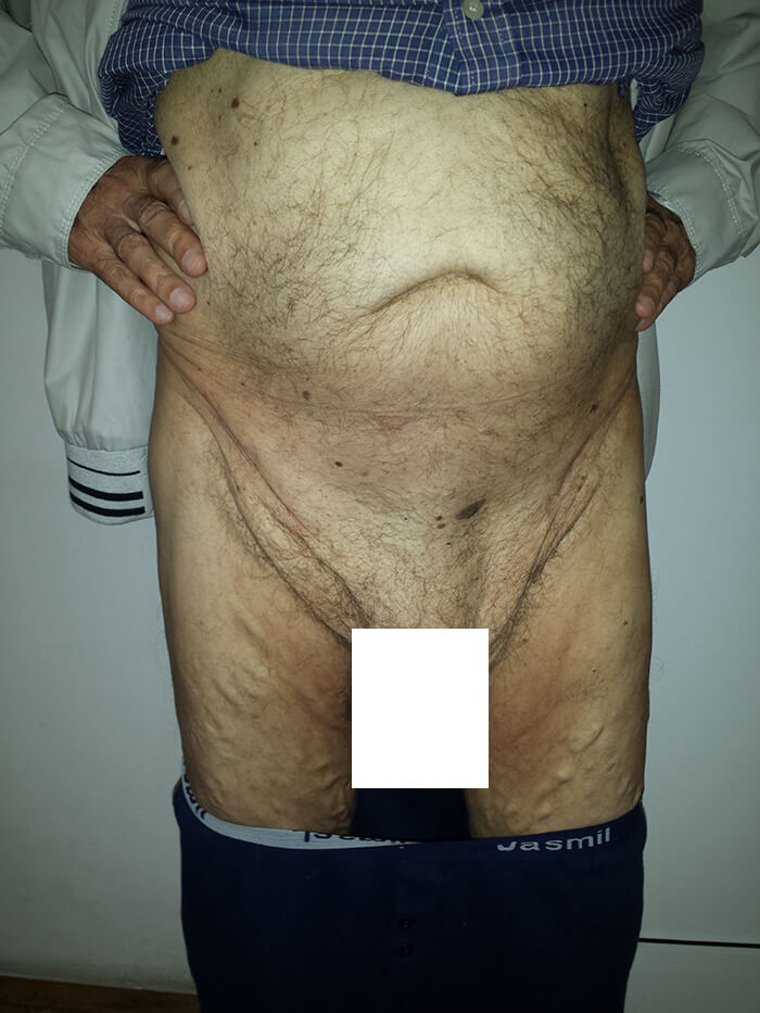
Pictures of different hernias of the abdominal wall
(patients operated on by dr Žuvela)
HERNIA CENTAR ŽUVELA
Films by dr Žuvela – hernia surgery

Operation of a patient with an inguinoscrotal hernia according to the principles of ambulatory surgery – Lichtenstein technique under local anesthesia
Operation of 2 patients with inguinoscrotal hernia according to the principles of ambulatory surgery – Lichtenstein technique under local anesthesia: the surgeon operates on the surgeon, and then the surgeon who was operated on operates on his patient
Operation of a patient with incarcerated umbilical hernia according to the principles of ambulatory surgery – “Open preperitoneal flat mesh technique” under local anesthesia
Operation of a patient with inguinoscrotal hernia “Modified Rives technique performed through a direct inguinal approach
